Weight-bearing MRI, also known as Axial-loaded MRI, simulates a standing up position by applying compression to the lumbar spine. The imaging captures how the patient actually feels while standing, and takes into account the effect that gravity has on a patient’s spine. The MRI technician attaches the DynaWell compression device to the patient prior to entering the open MRI and applies 50% of the patient’s body weight in compression. This closes the gaps in the L-spine thereby revealing herniation and bulges that did not present as significantly when the patient was in a relaxed position. Adding 50% compression does not create false positives of medical conditions that do not exist.
Weight-bearing MRI provides the highest quality imaging available for the L-spine and can influence physician treatment decisions. Most importantly, it gives the physician more insight into the true nature of the patient’s condition, and gives the patient more comfort in knowing their doctor has found their source of pain that previously went undetected.
Furthermore, weight-bearing MRIs are currently used around the country even though M1 Imaging is the first facility to bring this groundbreaking technology to Michigan. The benefits of weight-bearing imaging have not been disputed and serve as direct evidence in court cases.
LET US ANSWER ALL YOUR QUESTIONS
FAQ FOR MRI / Procedures
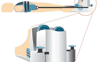 What is a weight-bearing MRI and how does it work?
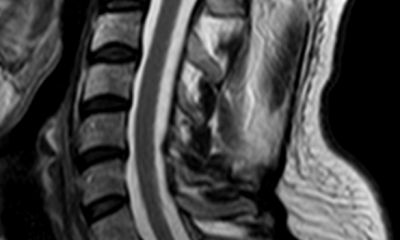
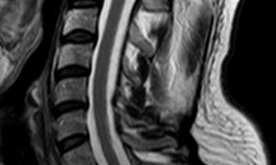 What is Flexion and Extension MRI for C-Spine?
Flexion and Extension MRI allows the physician to see the patient’s neck not only while it is in a supine position but also while the neck is bent up and down. Many times patients with whiplash or other neck conditions are in greater pain while they are flexing and extending their neck. M1 Imaging is able to capture these discords while other MRI centers in the area do not provide this technique. The patient benefits from having better image data and it does not take very long to add flexion and extension to the patient’s scan time.
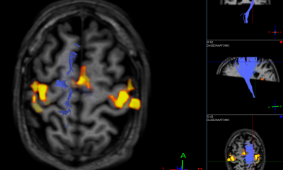
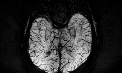 How is the M1 Imaging protocol different than most MRI centers for Traumatic Brain Injuries?
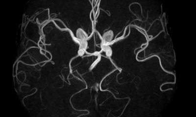
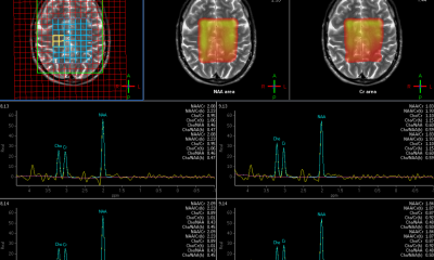
M1 Imaging uses a specialized protocol called Susceptibility Weighted Image (SWI). The SWI image is possible through the use of algorithms that differentiate the densities of adjacent tissues. While the signal density forms the basis of all MR scans, the SWI scan is 3-to-6 times more sensitive as it accounts for the susceptibility of all brain elements, including hemorrhages – hence the name susceptibility-weighted image. This differs from the more common DTI protocol. DTI works by measuring the diffusion of water through tissue. With DTI, measurements are isolated to identify the preferred direction of flow, which allows for the isolation of neural tracts from the brain’s white matter. The SWI image shows iron concentrations in the brain and can detect a brain hemorrhage even when the general brain scan appears negative.
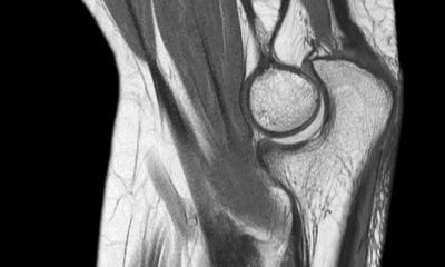
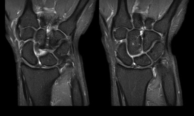
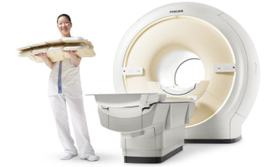 Does M1 Imaging have better dynamic joint imaging than most MRI facilities?
Yes, M1 Imaging uses High Field coils that specifically target the joints and provide much better images than competitor MRIs. Our imaging of shoulders, elbows, hips, knees, ankles, and feet provide a greater level of detail and much higher resolution than most facilities can offer.
 What is M1’s reporting turn-around time and are the reports accessible online?
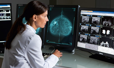
Dr. Chintan Desai, the chief radiologist for M1 Imaging, reads reports on a daily basis and our office will fax your patient’s report within 24-48 hours. Physicians are given a username and password to log into their own online database where they can see all their patient’s reports in one place, as well as the key images and prior reports. Physician staff can download a PDF version of the report onto a computer hard drive and upload it into a patient’s file.
 Do you offer ACR portal access?
M1 allows its physicians to access the American College of Radiology website to guide appropriateness of MRI ordering. The website will help doctors determine which MRIs are needed based on a set of symptom criteria. This way, physicians can be sure they are ordering the right MRI protocol for their patient’s condition.
 Does M1 Imaging charge extra for these specialized MRIs?
No, M1 Imaging provides exceptional value to patients. We bill at 60% of the national average, and we do not charge additional fees for the weight-bearing, flexion and extension, and TBI protocol. We take almost all insurance including BCBS, Medicare and Medicaid.
FAQ FOR Patients
 What’s an MRI?
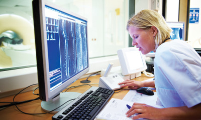
MRIs or Magnetic Resonance Images let doctors see inside your body to identify a wide variety of possible medical conditions—all without exposure to X-rays. Instead, an MRI uses a powerful magnet, radio waves, and special coils to detect electrical signals from your body. A computer then processes this information to create detailed images our doctors can use to find out what’s wrong with you or a family member.
 Why is M1 the right choice for an MRI?
At M1 Imaging Center we ensure our doctors and staff have access to the best imaging equipment available and help deliver the best experience possible to our patients. More accurate scans mean less repeat exams for you! Also, we are conveniently located with parking right in front of our building. We provide you with a CD after your MRI which you can then take with you to your doctor. We can also provide transportation if you are unable to drive. Just call our office to (248) 268-2119.
 How do I schedule an MRI at M1 Imaging Center?
In most cases, your doctor can do that for you. However, M1 will be in touch with you to confirm your appointment. If you have questions about getting an MRI at M1, be sure to contact us at (248) 268-2119.
 What kind of technology do you use?
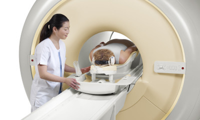
M1 Imaging Center uses a new Siemens Ingenia 1.5T open MRI system that offers vastly improved performance in three key areas: image quality, exam speed, and patient comfort. We are also the ONLY facility in the entire state of Michigan with the DynaWell L-Spine compression device for axial loaded weight-bearing MRIs so that you have the most accurate and reliable scan available in the market today.
 Do MRIs hurt?
MR imaging is painless and much quicker than you might think, especially with our new Philips Ingenia 1.5T MRI technology. It has a wide opening, so it’s much less confining than older MR systems and a lot more comfortable for more patients. Since it’s fast and accurate, you’ll be done quickly and it’s less likely your doctor will have to run any follow-up scans. If you are having a compression-based L-Spine MRI, you may experience the same discomfort as when you are standing even though you will be laying down.
 Is there any risk?
MRIs use no harmful radiation and there are no known health side effects. MRI studies are very low-risk procedures for most patients. However, an MRI isn’t for everyone. Be sure to inform your physician if you have a pacemaker, aneurysm clips in the brain, a shunt with telesensor, inner ear implants, metal fragments in one or both eyes, implanted spinal cord stimulators, or if you’re pregnant or breast feeding.
 How do I prepare for my MRI exam?
For many MRI exams, no special preparation is needed. For some, you may need to fast for 4-12 hours prior to the exam. Others require you to swallow a fluid, or to have an injection, that helps show what’s going on inside your body. Guidelines vary with the specific exam and also with the facility. You will be asked to remove all metal (earrings, watches, bobby pins, etc.) and credit cards. You may be asked to wear a gown during the exam or wear loose-fitting clothing with no metal fasteners, zippers, or buttons.
 Does the scanner make a lot of noise?
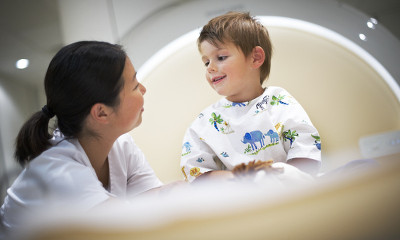
The magnet makes a slight rapping sound as images are being taken. In between scans the machine is quiet. Your MRI technologist will provide you with hearing protection, but you can still hear the technologist if he or she speaks to you during the exam.
 Will I get claustrophobic?
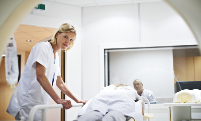
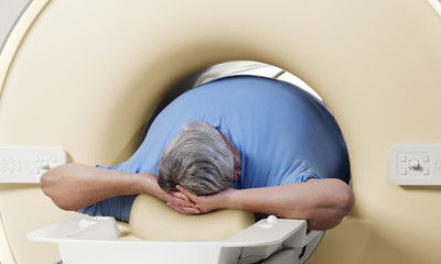
M1 Imaging Center offers state-of-the art technology and an “open” MRI system that you can feel good about. It’s based around a wide-bore 70cm magnet, and has a much more open feel than conventional MR systems. The open system allows us to image patients with diverse sizes (up to 550 lbs), ages, and physical conditions. In fact, most scans can be performed with your head and feet entirely out of the opening. You’ll probably be very comfortable as you lie on the padded table.
 Will I be alone?
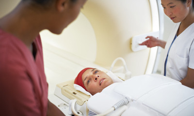
You’ll be in contact with one of our technologists at all times. Even when he or she is not in the MRI room, you will be able to talk to him or her by intercom. In some cases a family member is welcome to stay in the room with you during your scan.
 Will I have to hold still the whole time?
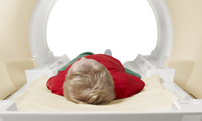
We’ll get the highest quality results if you hold still during the exam. The technologists will guide you and let you know when you can move between scans.
 How long will the exam take?
With our advanced MRI technology, we can perform routine exams of the brain, spine, knee, ankle, and liver quickly and easily, some cases in less than 8 minutes – with superb image quality. More complex vascular, cardiac, and musculoskeletal studies can last from 20 to 40 minutes. You should allow extra time in case the exam lasts longer than expected.
FAQ FOR Providers
 Why should I refer patients to M1 Imaging Center for an MRI?
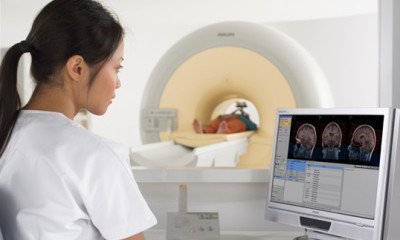
You can refer your patients to M1 with confidence because we have invested in the Philips Ingenia 1.5T, the first-ever digital broadband magnetic resonance imaging system. It’s an advanced MR scanner that provides both consistently high-quality images and a high level of patient comfort. The Ingenia 1.5T delivers excellent signal-to-noise ratio, streamlines workflow, and provides an open patient experience.
We are also the ONLY facility in the entire state of Michigan with the DynaWell L-Spine compression device for axial loaded weight-bearing MRIs so that you have the most accurate and reliable scan available in the market today. Research has shown that weight-bearing MRI’s increase detection results and help doctors make better treatment choices for their patients.
Our Specialties:
- Weight-bearing MRI for L-Spine
- Flexion & Extension MRI for C-Spine
- Traumatic Brain Injury Protocol with Susceptibility-Weighted Imaging (SWI)
- High Field Coils for Targeted Joint MRI
 How will my patients feel about your technology?
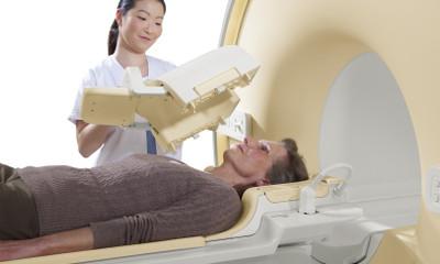
M1 Imaging Center offers state-of-the art technology and a mobile “open” MRI system that your patients can feel good about. It’s based around a wide-bore 70cm magnet, and has a much more open feel than conventional MR systems. The open system allows us to image patients with diverse sizes (up to 550 lbs), ages, and physical conditions. In fact, most scans can be performed with their head and feet entirely out of the opening, so your patients are likely to feel less claustrophobic and more relaxed about getting an MR scan. Additionally, our DynaWell L-Spine compression device simulates a stand-up position while lying down.
 What are the benefits?
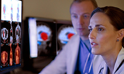
Excellent images – The Philips Ingenia 1.5T is the first digital broadband system ever. It incorporates dStream architecture that digitizes the signal directly in the MR coil, substantially improving the signal-to-noise ratio, resulting in exceptional image resolution. The DynaWell L-Spine compression device can provide 60-70% more data for patients with symptoms of Sciatica and Neurogenic Claudication.
Fast exams – With our advanced MRI technology, we can perform routine exams of the brain, spine, knee and as well as organs and extremities quickly and easily, some cases in less than 8 minutes – with superb image quality.
More complex vascular, cardiac, and musculoskeletal studies can last from 20 to 40 minutes. The Ingenia 1.5T has innovative technologies which speed up exams and increase productivity. It simplifies patient positioning and coil handling for our technologists, so patients can be scanned from head to toe across the entire 55cm field of view in less time than current MR systems.Fewer repeat exams – The Ingenia 1.5T gives our radiologists and referring physicians access to the clear, detailed MR images they need to help make informed diagnoses, resulting in fast, more comfortable exams and fewer repeat exams for our patients. Published studies in, among others “Spine”, and ongoing studies verify that the DynaWell® methodology is valuable in lumbar spine pathology (spinal stenosis, sciatica, disc herniation, synovial cysts) and hip, knee and ankle pathology. With DynaWell®, clinical studies show that it is possible to achieve a more specific and valid diagnosis, compared with regular non-loaded PRP.
 What diagnostic imaging applications does M1 Imaging Center handle?
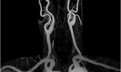
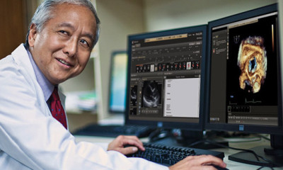
Our Philips Ingenia 1.5T was designed to deliver precise, detailed MR images to radiologists and referring physicians who need to diagnose many different anatomical and structural problems in the body.
 Does M1 take health insurance? How much does a DynaWell compression-based MRI cost?
Yes, M1 Imaging accepts mostly all healthcare plans including BCBSM and Medicare. The addition of the DynaWell compression device does not increase the cost of the L-Spine MRI.
 How do I contact M1 Imaging Center?
If you have questions about scheduling, or the benefits of M1 Imaging Center for all your MRI needs, please contact us using for form below.

