FAQ related to
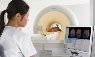 Why should I refer patients to M1 Imaging Center for an MRI?You can refer your patients to M1 with confidence because we have invested in the Philips Ingenia 1.5T, the first-ever... Read More
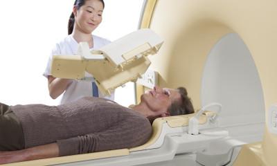 How will my patients feel about your technology?M1 Imaging Center offers state-of-the art technology and a mobile “open” MRI system that your patients can feel good about.... Read More
 What are the benefits?Excellent images – The Philips Ingenia 1.5T is the first digital broadband system ever. It incorporates dStream architecture that digitizes... Read More
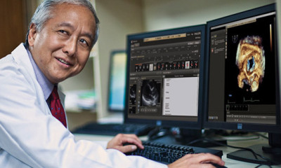 What diagnostic imaging applications does M1 Imaging Center handle?Our Philips Ingenia 1.5T was designed to deliver precise, detailed MR images to radiologists and referring physicians who need to... Read More
 Does M1 take health insurance? How much does a DynaWell compression-based MRI cost?Yes, M1 Imaging accepts mostly all healthcare plans including BCBSM and Medicare. The addition of the DynaWell compression device does... Read More
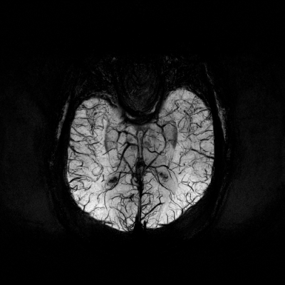
 What’s an MRI?MRIs or Magnetic Resonance Images let doctors see inside your body to identify a wide variety of possible medical conditions—all... Read More
 Why is M1 the right choice for an MRI?At M1 Imaging Center we ensure our doctors and staff have access to the best imaging equipment available and help... Read More
 How do I schedule an MRI at M1 Imaging Center?In most cases, your doctor can do that for you. However, M1 will be in touch with you to confirm... Read More
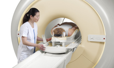 What kind of technology do you use?M1 Imaging Center uses a new Siemens Ingenia 1.5T open MRI system that offers vastly improved performance in three key... Read More
 Do MRIs hurt?MR imaging is painless and much quicker than you might think, especially with our new Philips Ingenia 1.5T MRI technology.... Read More
 Is there any risk?MRIs use no harmful radiation and there are no known health side effects. MRI studies are very low-risk procedures for... Read More
 How do I prepare for my MRI exam?For many MRI exams, no special preparation is needed. For some, you may need to fast for 4-12 hours prior... Read More
 Does the scanner make a lot of noise?The magnet makes a slight rapping sound as images are being taken. In between scans the machine is quiet. Your... Read More
 Will I get claustrophobic?M1 Imaging Center offers state-of-the art technology and an “open” MRI system that you can feel good about. It’s based... Read More
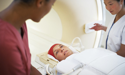 Will I be alone?You’ll be in contact with one of our technologists at all times. Even when he or she is not in... Read More
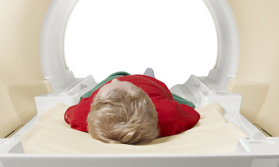 Will I have to hold still the whole time?We’ll get the highest quality results if you hold still during the exam. The technologists will guide you and let... Read More
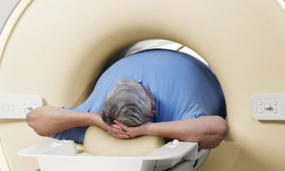 How long will the exam take?With our advanced MRI technology, we can perform routine exams of the brain, spine, knee, ankle, and liver quickly and... Read More
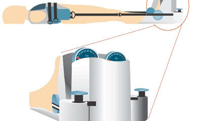 What is a weight-bearing MRI and how does it work?Weight-bearing MRI, also known as Axial-loaded MRI, simulates a standing up position by applying compression to the lumbar spine. The... Read More
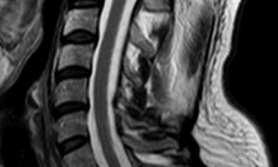 What is Flexion and Extension MRI for C-Spine?Flexion and Extension MRI allows the physician to see the patient’s neck not only while it is in a supine... Read More
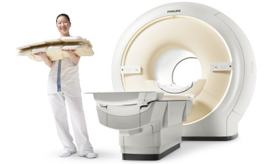 Does M1 Imaging have better dynamic joint imaging than most MRI facilities?Yes, M1 Imaging uses High Field coils that specifically target the joints and provide much better images than competitor MRIs.... Read More
 What is M1’s reporting turn-around time and are the reports accessible online?Dr. Chintan Desai, the chief radiologist for M1 Imaging, reads reports on a daily basis and our office will fax... Read More
 Do you offer ACR portal access?M1 allows its physicians to access the American College of Radiology website to guide appropriateness of MRI ordering. The website... Read More
 Does M1 Imaging charge extra for these specialized MRIs?No, M1 Imaging provides exceptional value to patients. We bill at 60% of the national average, and we do not... Read More
 How do I contact M1 Imaging Center?If you have questions about scheduling, or the benefits of M1 Imaging Center for all your MRI needs, please contact... Read More